
Images
NLST CT Images
Collection
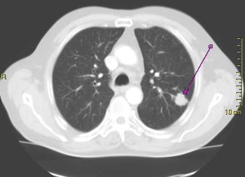
The NLST low-dose helical CT scan image collection comes from over 25,000 people who were randomly assigned to the CT arm of NLST. The CT arm protocol was for three annual helical CT exams to screen for lung cancer: one at baseline (T0) and two more on the first and second anniversaries of randomization (T1 and T2). Only images from CT screening exams are available; images from follow-up scans (with higher radiation dose and image quality) are not in the collection. For each screening exam, the image collection contains (on average) one localizer image and 2 to 3 axial reconstructions of a single helical CT scan of the chest. Approximately 200,000 image series from 75,000 CT exams in 25,000 people are available.
CT images do not contain radiologist's findings. Findings detected in trial screening can be found in the Spiral CT Abnormalities or Spiral CT Comparison Read Abnormalities datasets. Coordinates of findings on CT screens were not collected by the NLST trial.
Lung Cancer Selection
For investigator convenience, a subset of participants have been made available for direct download through an approved Cancer Data Access System (CDAS) project (~28,000 images from ~3,700 participants totaling ~870 GB). The Lung Cancer Selection provides investigators with a set of images and data that are designed to provide the participants with lung cancers (screen-detected and otherwise) and a cross-section of control participants at a size that is manageable for download. Participants within the Lung Cancer Selection are divided into equally-sized training and testing sets designed to have an even distribution of cancers, nodule positive screens, and nodule negative screens. In detail, the Lung Cancer Selection includes:
- All CT images from all participants with screen-detected cancer (N = 623).
- All CT images from all participants with interval or post-screening lung cancer (N = 438).
- All CT images from a random selection of participants with at least one nodule-positive screen (N = 2120).
- All CT images from a random selection of participants with negative screens in all 3 study years (N = 527).
Coronary Artery Calcium (CAC) Selection
Also available for investigator convenience is a subset of participants focused on CAC investigation (~26,000 images from ~3,800 participants totaling ~1,260 GB). Participants in the CAC selection are divided into equally sized training and testing sets designed to have an even distribution of CAC cases and controls. CT images that overlap with the Lung Cancer selection are partitioned into separate batches so that non-overlapping images can be downloaded with ease.
- Participants with CT images showing CAC (N = ~2400)
- Participants with CT images that do not show evidence of CAC (N = ~1400)
Custom Selections
Investigators may opt to select their own custom populations with the assistance of CDAS staff to best suit their research needs. Custom selections are currently only available via hard drive and are not able to be downloaded from the CDAS website.
How to Access CT Images
CT images are available publicly and can be downloaded from The Cancer Imaging Archive or NCI Imaging Data Commons:
- TCIA: see the TCIA NLST collection page for access to the CT images in DICOM format
- IDC: open NLST collection in the IDC portal to access CT images in DICOM format
- These images can also be obtained through this website either via hard drive delivery or directly, for selected subsets, including the Lung Cancer and Coronary Artery Calcification selections.
The complete collection of CT images is comprised of more than 20 million files requiring over 10TB.
| Group | Participants | Localizer Images | Axial Images | Terabytes |
|---|---|---|---|---|
| Screen Detected Lung Cancer with Image in Same Study Year | 622 | 1,035 | 2,244 | 0.17 |
| Lung Cancer with Image | 436 | 1,000 | 2,104 | 0.16 |
| Nodule positive or screen positive with image in Same Study Year | 9,470 | 24,483 | 56,423 | 3.91 |
| 3 Negative Screens with 3 Images | 12,548 | 34,452 | 65,197 | 4.85 |
| Other Participants | 3,162 | 4,206 | 10,493 | 0.96 |
NLST Pathology Images

Collection
The NLST pathology images come from ~450 lung cancer patients in the Lung Screening Study (LSS) subcomponent of NLST. They are images of H&E-stained slides that were obtained as part of a pathology specimen collection to construct tissue microarrays (TMAs). The ~1250 H&E slides (and corresponding images) each came from blocks of lung tissue that were resected during diagnosis and treatment of lung cancer and preserved by pathology labs.
A pathologist reviewed each image to identify histologies of interest for tissue sampling and inclusion in the TMAs. Each region of interest (ROI) was digitally annotated, and the histologies were recorded on case report forms. Data from the case report forms may be obtained by investigators. Annotated pathology images were not retained and are therefore not available.
How to Access Pathology Images
Digital pathology images are available publicly and can be downloaded from The Cancer Imaging Archive or NCI Imaging Data Commons:
- TCIA: see the TCIA NLST collection page for access to the pathology images in SVS format
- IDC: open NLST collection in IDC portal to access pathology images in DICOM format
You may also request the pathology images by applying for an NLST project through CDAS. Upon project approval, investigators can download the entire collection of NLST pathology images through CDAS or may opt to receive the pathology images either via hard drive delivery or direct download.
If you have any questions please post at: https://cdas.cancer.gov/contact/nlst/External Sources
- Imaging Data Commons (IDC) - CT and Pathology images are available without login, and may be viewed online and downloaded. Questions related to the use of this service should be directed to https://discourse.canceridc.dev/.
- The Cancer Imaging Archive (TCIA) - CT and Pathology images may be downloaded. CT images are available in whole or in batches of 2,500 participants each. Questions related to the use of this service should be directed to help@cancerimagingarchive.net.
- American College of Radiology Imaging Network (ECOG-ACRIN). The x-ray images collected at ACRIN centers can be requested by contacting ACRIN (ECOG-ACRIN). Availability of additional ACRIN x-ray images can be requested by selecting "Contact Us". By completing the form and choosing the subject of "Data Sharing", the pertinent ECOG-ACRIN staff members (including the Data Sharing Administrator at Biostatistics Center) will be notified of the request and will respond to the investigator within 48 working hours. [Note: X-ray images collected at LSS centers were not digitized and are therefore not available.]